| Nipple | Slide 15 (H & E) |
| This slide demonstrates the different structures that form the nipple. |
|---|
|
This is a low magnification of a lactiferous duct illustrating the structure.
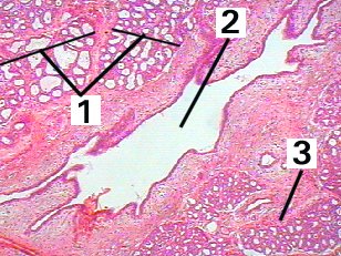
Fig 15-001 |
|
This is a very high magnification of the epithelium of a lactiferous duct.
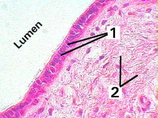
Fig 15-002 |
|
This is a very low magnification of the nipple demonstrating the structure.
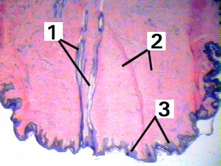
Fig 15-003 |
|
This is a higher magnification of a lactiferous sinus and surrounding connective tissue.
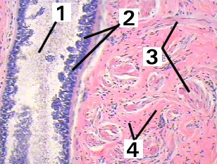
Fig 15-004 |
| Memorandum | ||
|---|---|---|
| Clinical case | Fig 15-001 | Fig 15-002 |
| Fig 15-003 | Fig 15-004 | |