| Mammary gland | Slide 57 (H & E) |
| This slide demonstrates the different structures that form the mammary gland. |
|---|
| Clinical Case |
|---|
|
A young female consults you because of a lump she has noticed in her left breast during self-examination. Her maternal grandmother had died of breast cancer.
Physical examination of the breasts reveals a hard nodule in the upper outer quadrant of the right breast. The nipple and areolae appear normal with no discharge. Although a mammogram is negative, you decide to refer her to a surgeon. The subsequent histopathological report reveals an extensive ductal carcinoma in situ.
|
| Microscopy |
|---|
|
This is a low magnification of an inactive breast demonstrating the different structures.

Fig 57-001 |
|
This is a higher magnification of an inactive breast demonstrating the glandular tissue.
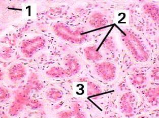
Fig 57-002 |
|
This is a low magnification of an active breast demonstrating the different structures.
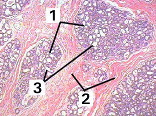
Fig 57-003 |
|
This is a very high magnification of the glandular alveoli of an active breast demonstrating the cells and tissue.
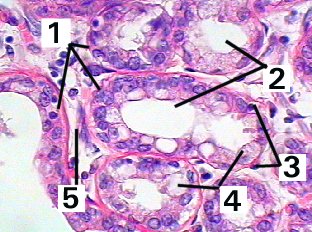
Fig 57-004 |
|
This is a low magnification of an example of carcinoma confined to the ducts of the mammary gland.
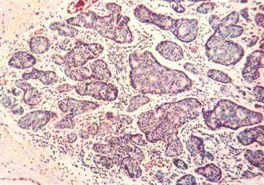
Fig Curran1208 |
|
This is a high magnification of an example of carcinoma confined to the ducts of the mammary gland.
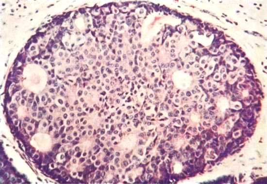
Fig Curran1209 |
| Memorandum | ||
|---|---|---|
| Clinical case | Fig 57-001 | Fig 57-002 |
| Fig 57-003 | Fig 57-004 | |