| Spleen | Slide 64 (H & E) |
| This slide demonstrates the different structures of the spleen. |
|---|
| Clinical Case |
|---|
|
While on duty in the casualty ward, you receive an emergency call from the sister in the day ward. A patient you admitted earlier the day for observation after a car accident was going into shock. The skin of the left hypochondrial area was severely bruised by the seat belt and because you were worried that he may have suffered a deceleration injury to the internal organs, you decided to have him observed for 24 hours.
When you see the patient in the ward, he is anxious and presents with tachycardia and hypotension, all signs of shock due to acute loss of blood. Suspecting a ruptured spleen, you refer him to surgery for an emergency laparotomy.
|
| Microscopy |
|---|
|
This is a low magnification of the spleen demonstrating the capsule and white and red pulps.

Fig 64-001 |
|
This is a magnification of the white and red pulp of the spleen.
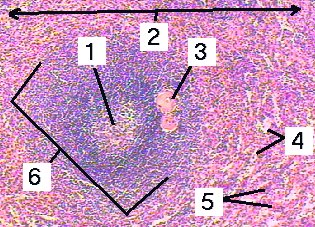
Fig 64-002 |
|
This is a very high magnification of the red pulp of the spleen demonstrating the different structures.

Fig 64-003 |
|
This is a very high magnification of the white pulp demonstrating the different structures.

Fig 64-004 |
|
This is a very high magnification of the structure of the peri-arterial lymphoid sheaths.

Fig 64-005 |
|
This is a very high magnification of the spleen stained with silver to demonstrate the reticular fibers around and in between the sinusoids.

Fig 64-006 |
| Memorandum | |||
|---|---|---|---|
| Clinical Case | Fig 64-001 | Fig 64-002 | Fig 64-003 |
| Fig 64-004 | Fig 64-005 | Fig 64-006 | |
© mei mmi marius loots