| Ileum | Slide 41 |
| The following five figures demonstrate the different structures of the ileum. |
|---|
| Clinical Case |
|---|
|
A 25-year old male student presents with fever, abdominal pain, diarrhea and complains of fatigue. He also says that he has lost weight.
Physical examination reveals right lower quadrant tenderness with a fullness reflecting adherent loops of the small bowel. A laparotomy showed the appearance of the terminal ileum to be hyperemic and boggy, with mesentery and mesenteric lymph nodes swollen and reddened, although the bowel wall was pliable. A diagnosis of Crohn's disease is made.
|
| Microscopy |
|---|
|
This is a very low magnification of a section through the ileum demonstrating the different layers and structures.
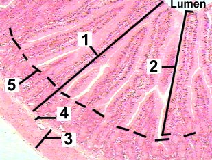
Fig 41-001 |
|
This preparation is a higher magnification of the ileum demonstrating structures of the mucosa.
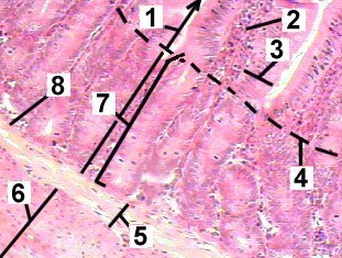
Fig 41-002 |
|
This is a very high magnification of the ileum epithelium.
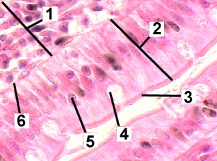
Fig 41-003 |
|
A very high magnification of the outer layers of the ileum.
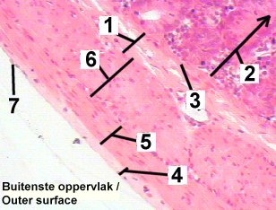
Fig 41-004 |
|
This figure is a high magnification of a Peyer's patch in the ileum.
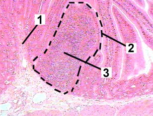
Fig 41-005 |
| Memorandum | ||
|---|---|---|
| Clinical Case | Fig 41-001 | Fig 41-002 |
| Fig 41-003 | Fig 41-004 | Fig 41-005 |