| Pancreas | Slide 50 (H & E) |
| Clinical Case |
|---|
|
A patient with a known possible predisposition to pancreatitis presents with severe and constant abdominal pain, nausea, tachycardia, and abnormal findings on abdominal examination.
Laboratory studies reveal leukocytosis, an abnormal appearance on x-rays of the abdomen and chest and hyperglycemia. The diagnosis is confirmed by the finding of an elevated level of serum amylase.
|
| Microscopy |
|---|
| The following four figures demonstrate the different structures of the pancreas. |
|---|
|
This is a low magnification of the pancreas demonstrating the lobuli and septa.

Fig 50-001 |
|
This is a low magnification of the pancreas demonstrating the islands of Langerhans.
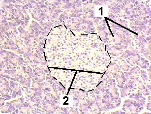
Fig 50-002 |
|
This is a very high magnification of the pancreas demonstrating the fine structure of the island of Langerhans (endocrine pancreas).
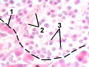
Fig 50-003 |
|
This is a very high magnification of the pancreas demonstrating the fine structure of the pancreatic acini of the exocrine pancreas.
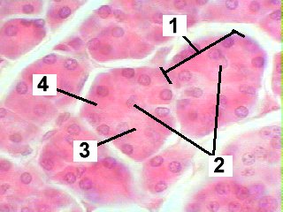
Fig 50-004 |
| Memorandum | ||
|---|---|---|
| Clinical Case | Fig 50-001 | Fig 50-002 |
| Fig 50-003 | Fig 50-004 | |