The Circulatory System

  • The blood vascular system comprises three parts, namely the arterial system, microcirculation and venous system.
  • The arterial system is composed of large elastic arteries, middle sized or muscular arteries and arterioles.
  • The microcirculation encompasses capillaries into which the arterioles drain and which in turn drain into sinusoids and venules forming networks in the tissues for the exchange of gases, fluids, metabolites and waste.
  • The venous system comprises small muscular venules, small to middle sized veins and large veins.
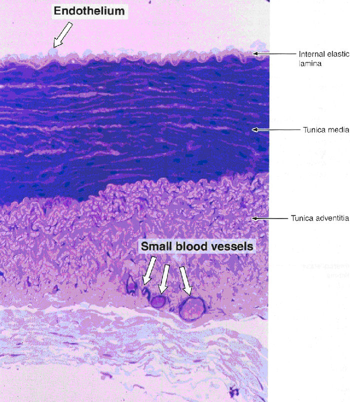
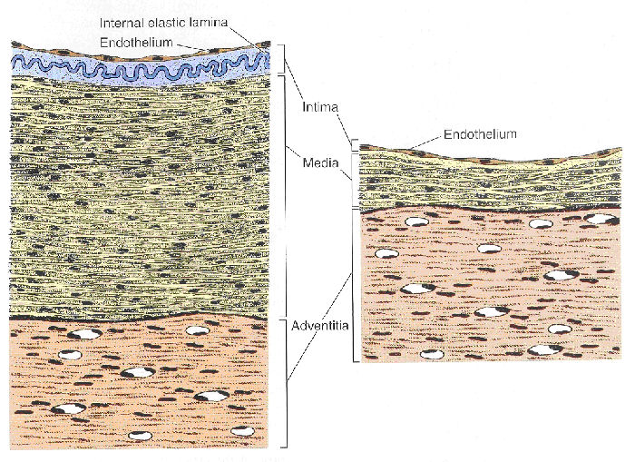
  • Morphologically the walls of all blood vessels have been defined into three layers or tunics down to arteriolar and venular level.
  • These tunics have been named from inside to outside as the tunica intima, tunica media and tunica adventitia.
  • The presence and thickness of the tunics vary according to the location and function of the blood vessel (Fig 11.15)
Fig 12.1

Fig 12.1 Ross and Romrell, p. 284
Fig 11.15

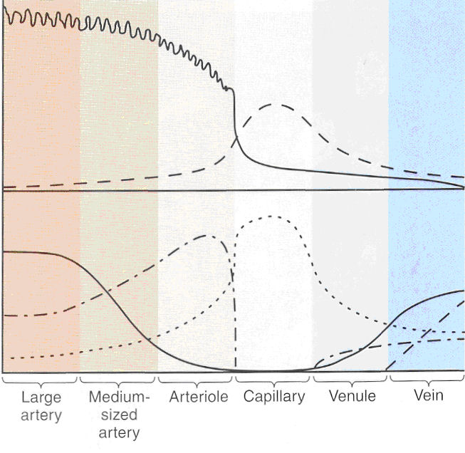
Fig 11.15 Junqueira and Carneiro p. 225

Graph showing the relationship between the characteristics of the blood circulation and the structure of the blood vessel. The arterial blood pressure and speed of flow decrease and become more constant as the distance from the heart increases. This decrease coincides with the reduction in the number of elastic fibers and an increase in the number of smooth muscle cells in the arteries. The graph illustrates the gradual changes in the structure of the vessels and their biophysical properties.

  1. The tunica intima
    • The tunica intima generally is composed of a single flattened endothelial layer lining the lumen of the blood vessel.
    • Beneath the tunica intima is the subendothelial layer of connective tissue, a few elastic fibers, fibroblasts and in some cases smooth muscle fibers.
    • The subendothelial layer is situated against the lamina elastica interna which is a fenestrated elastic membrane consisting of elastin.
  2. The tunica media
    • The tunica media consists principally of smooth muscle fibers with a little connective tissue between the smooth muscle fibers.
    • Surrounding the tunica media is a layer of elastic fibers, termed the lamina elastica externa.
  3. The tunica adventitia
    • The surrounding tunica adventitia is built up of connective tissue which blends with the surrounding connective tissue. The larger blood vessels are supplied by nutrient vessels called the vasa vasorum.
Fig 11.7

Junqueira and Carneiro p. 219

1. The arterial system

  • The arterial system is characterized by a more prominent tunica media in relation to the tunica intima and tunica adventitia.
  • The thickness of the wall of arteries is about as broad as the diameter of the lumen.

1.1 The large or elastic arteries

  • E.g., the aorta
  • The tunica intima forms about 20% of the wall thickness
  • The lamina elastic interna is a fenestrated elastic membrane.
  • The media is the thickest layer and consists of concentric fenestrated elastic membranes separated by collagen fibers and a few smooth muscle fibers.
  • A lamina elastica externa is not visible.
  • The adventitia contains a vasa vasorum.
Fig 11.14

Junqueira and Carneiro p. 223

1.2 The middle sized or muscular arteries

  • E.g., the femoral arteries
  • The intima is generally thrown into wavy folds
  • The lamina elastica interna is a prominent fenestrated elastic membrane consisting of elastin.
  • The media is the most prominent layer and consists mainly of smooth muscle fibers with a little connective tissue interspersed between.
  • The media is surrounded by a fairly thick lamina elastica externa.
  • The adventitia is generally thin but can sometimes be as thick as the media
Fig 11.13

Junqueira and Carneiro p. 223

1.3 The arterioles

Fig 11.9

Junqueira and Carneiro p. 220
Fig 11.12

Junqueira and Carneiro p. 222

2. The Venous System

  • The venous system is characterized by a more prominent tunica adventitia in relation to the tunica intima and tunica media
  • The walls of veins are thin relative to the diameter of the lumen
Fig 11.10

Junqueira and Carneiro p. 221

2.1 Large veins

  • E.g., the femoral veins
  • The intima of these veins are similar to that of middle sized veins except for a thicker subendothelial layer
  • The media of large and middle sized veins are also similar
  • The prominent and thickest layer is the adventitia which consists of bundles of collagen fibers with elastic fibers. Interspersed between the bundles of collagen fibers are bundles of smooth muscle fibers.
Fig 11.19

Junqueira and Carneiro p. 228

2.2 The middle sized veins

  • E.g., the vessels that collect the blood from the venules
  • Middle sized veins are characterized by a very thin intima that consists of little more than the endothelial layer
  • The larger veins have a small amount of subendothelial connective tissue
  • The lamina elastica interna is poorly developed
  • In smaller veins the media and adventitia are of the same width
  • The valves of these veins are formed by intimal folds
Fig 11.18 Muscular artery vs. Vein

Junqueira and Carneiro p. 227

3. The heart

  • The wall of the heart also contains three tunics similar to blood vessels
  • The tunica intima of the heart is called the endocardium
  • The tunica media is termed the myocardium
  • The tunica adventitia is called the epicardium
  • Beneath the intima is a dense fibro-elastic layer which becomes continuous with the perimysium of the cardiac muscle
  • The myocardium is the thickest layer of the heart and is composed of cardiac muscle
  • Mesothelium supported by a thin loose connective tissue forms the epicardium
Fig 8.1

Wheater p. 144

Key:
A - Coronary artery
Epi - Epicardium
M - Myocardium
E - Endocardium
P - Pericardium

Fig 8.5

Wheater p. 146

Key:
S - Fibroelastic supporting layers
F - Lamina Fibrosa

4. The lymphatic system

Is a drainage system which drains lymph from the extracellular space and returns it to the blood vascular system Lymphatic vessels have a similar structure to blood vessels The lymphatic vessels contain valves, although they are more numerous
Fig 8.22

Wheater p. 156

Key:
L -Lymphatic vessel
V - Venule

Fig 8.23

Wheater p. 156

Key:
V - Lymphatic vessel valve

back


© augustus 2007 marius loots