| The following three figures demonstrate the structure of secretory units secreting specific types of secrete. |
|---|
| Submandibular salivary gland | Slide 35 (H & E) |
|
A very high magnification of the submandibular gland demonstrating the structure and arrangement of the cells of a serous secretory unit.
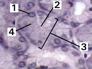
Fig 35-001 Wheater 3rd Ed p.91 or 4th Ed p. 95 |
| Pharyngeal tube (Eustachian tube) | Slide 39 (H & E) |
| The following two figures illustrate the different structures of mucinous and mixed glands. |
|---|
|
A very high magnification of the pharyngeal tube demonstrating the structure and arrangement of the cells of a mucinous secretory unit.

Fig 39-003 Wheater 3rd Ed p.91 or 4th Ed p. 95 |
|
A very high magnification of the pharyngeal tube demonstrating the structure and arrangement of the cells of a mixed secretory unit.

Fig 39-004 Wheater 3rd Ed p.91 or 4th Ed p. 95 |
| Memorandum | |||
|---|---|---|---|
| Clinical Case | Fig 35-001 | Fig 39-003 | Fig 39-004 |