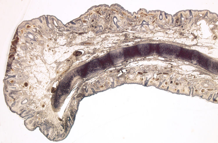
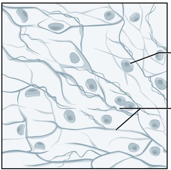
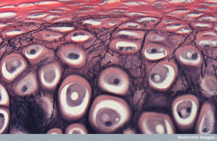
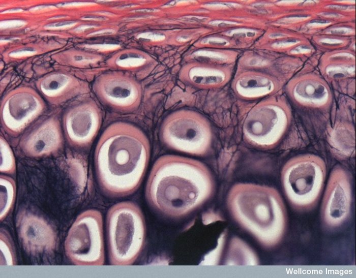
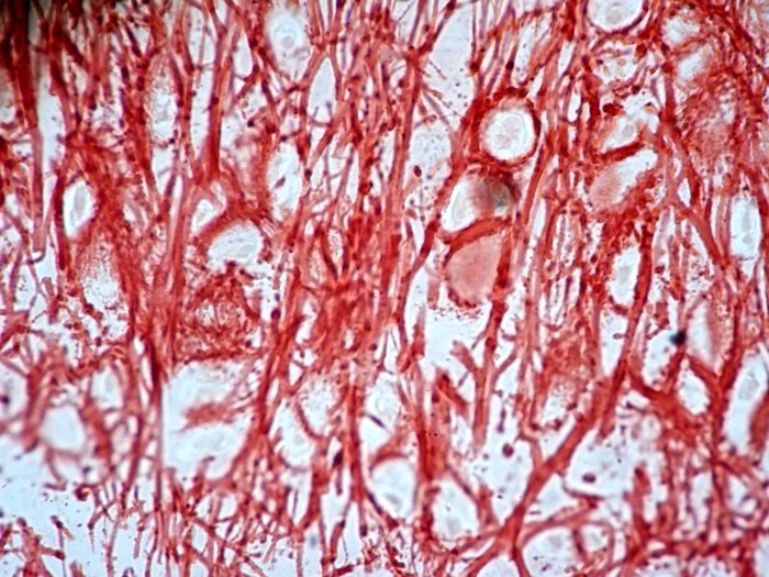
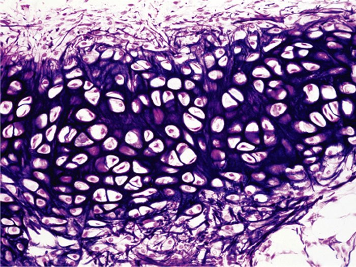
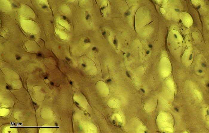
Slide 9: External ear
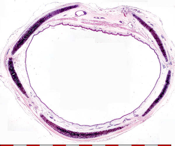
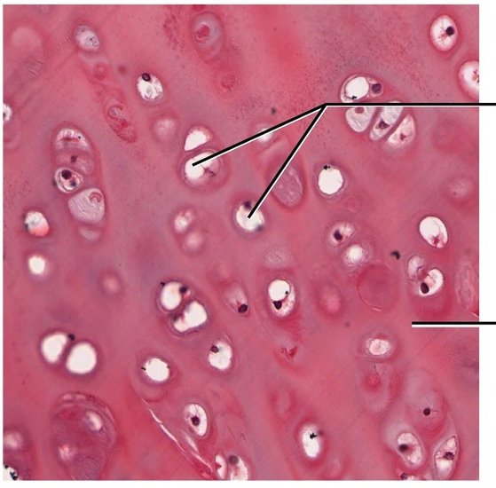
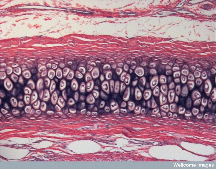
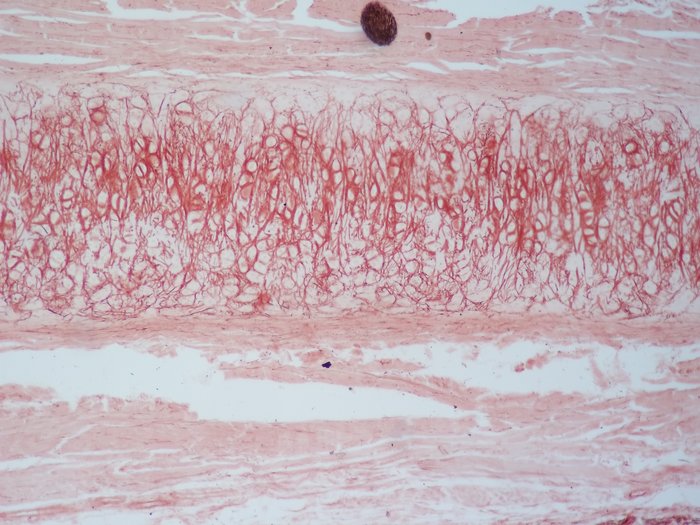
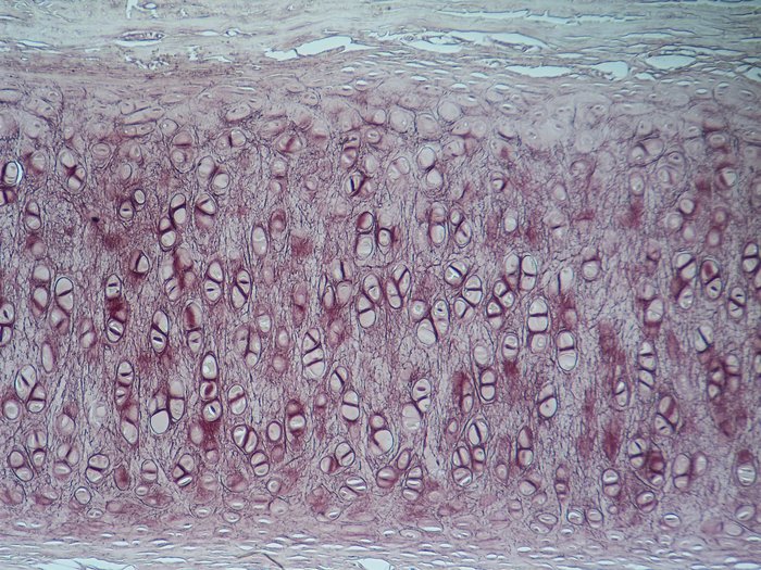
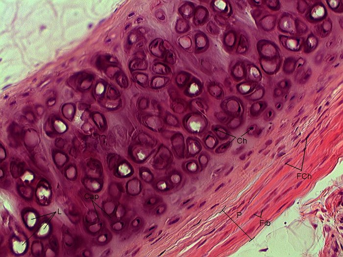
Slide 73: Trachea
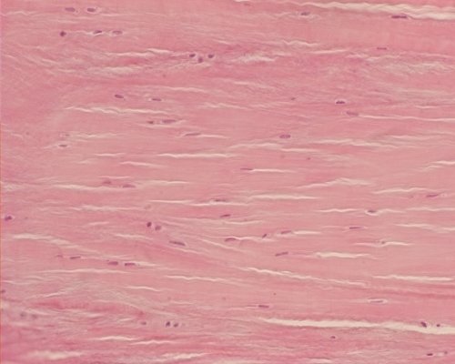
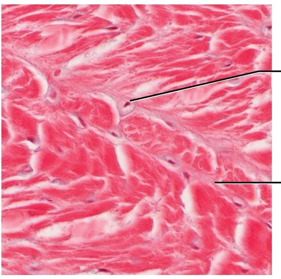
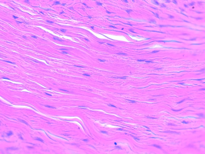
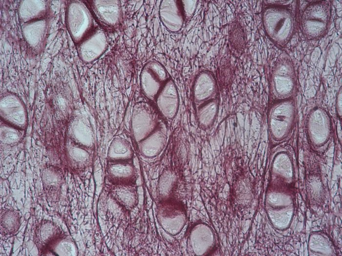
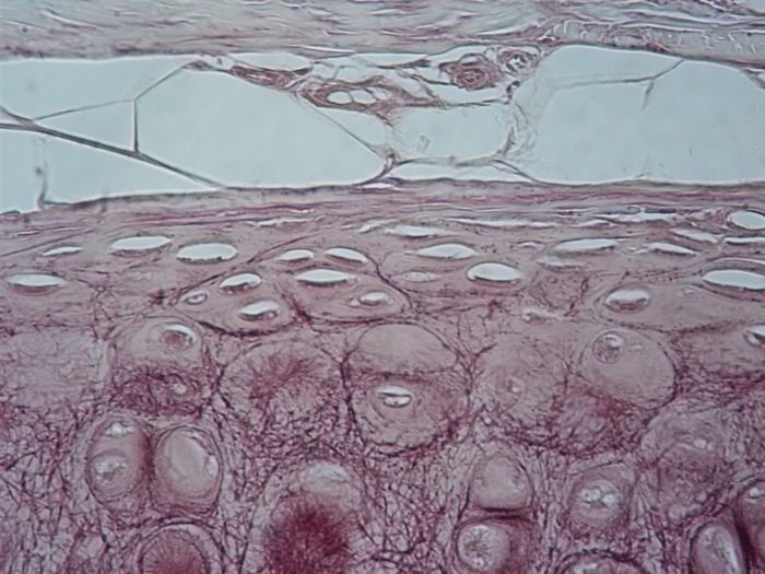
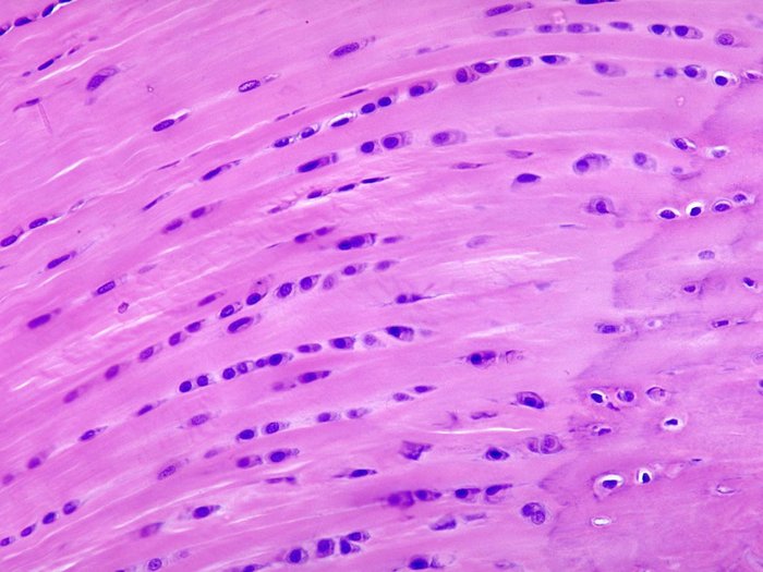
Slide 10: Fibro-cartilage
??

Slide 9: External ear
|
Slide 73: Trachea
|
Slide 10: Fibro-cartilage
|
|
|
|
|
|
|
|
|
|
|
|
|
|
|
??
|
|
|
|
|
|
|
|
|
|
|
|
|
|
|
|
|
|
|
|
|
|

|
|
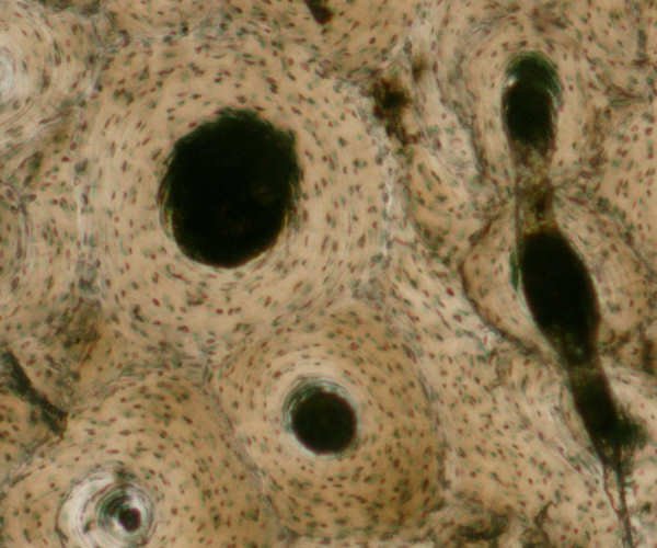
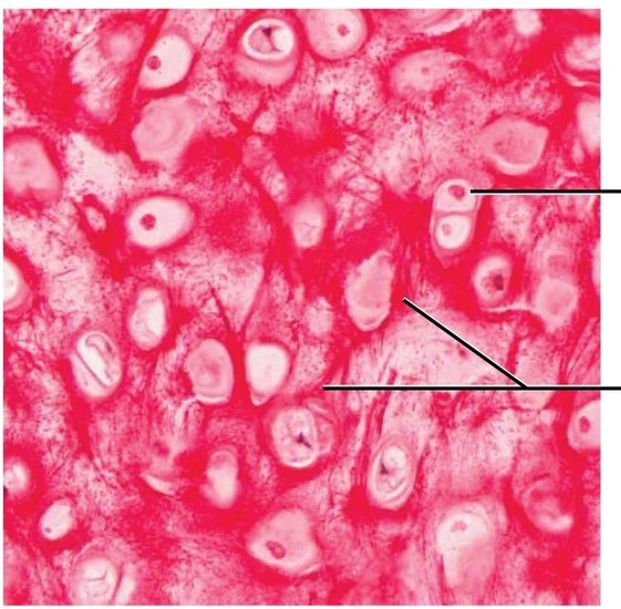
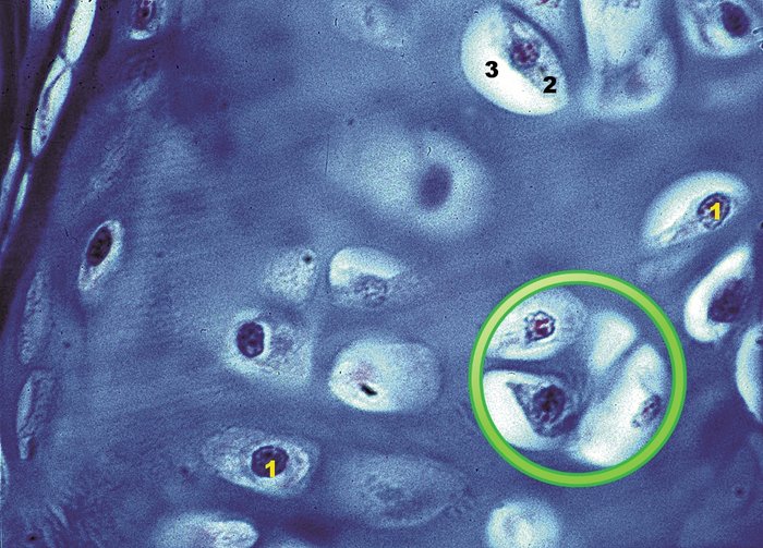
Slide 13: Cross section of Haversiona System
|
|

|
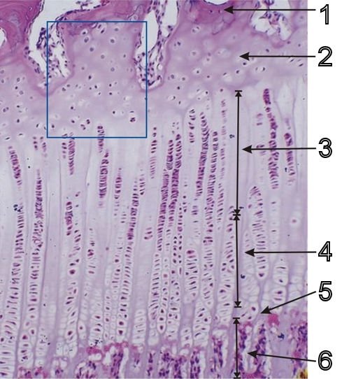
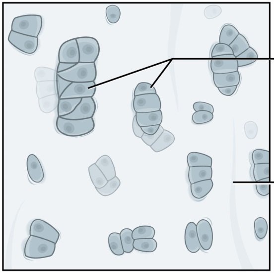
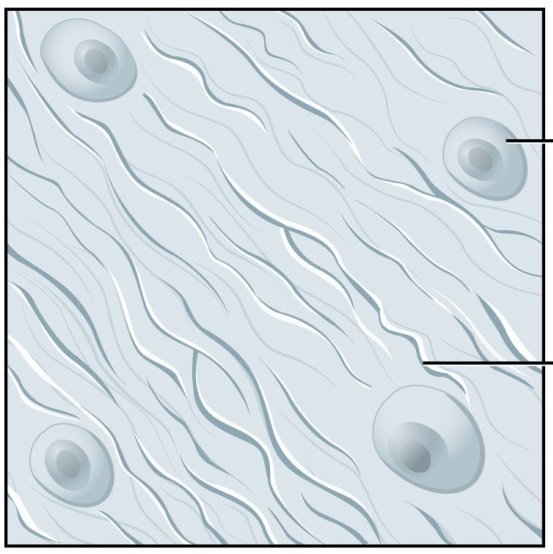
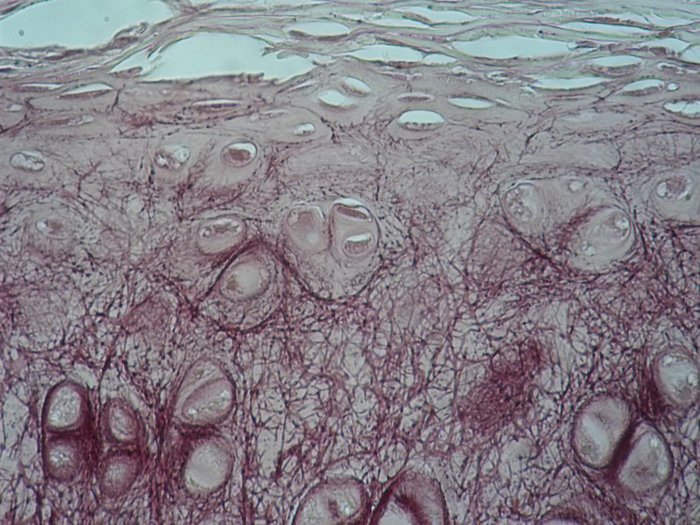
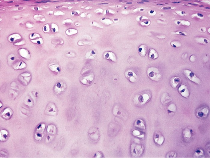
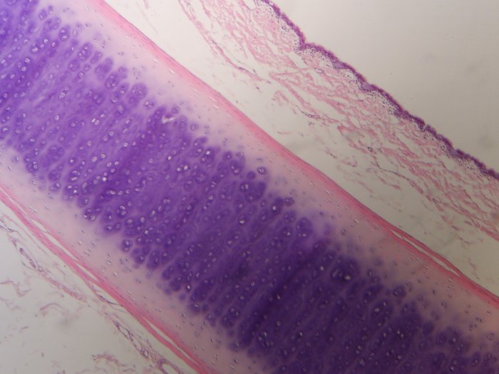
Annotate the zones and describe the changes taking place in each zone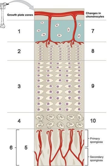
|
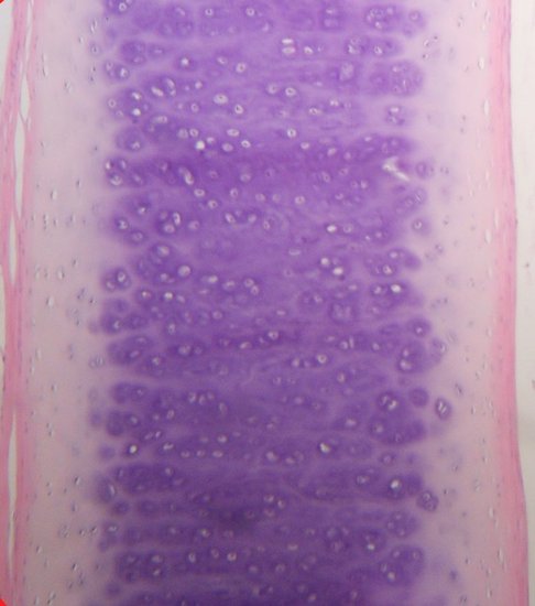
Annotate the zones and describe the changes taking place in each zone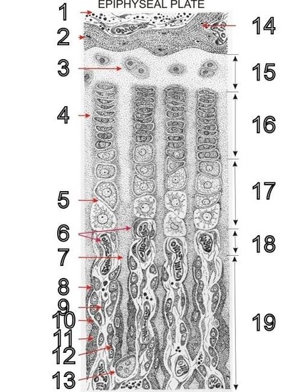
|
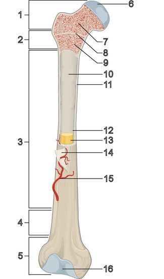
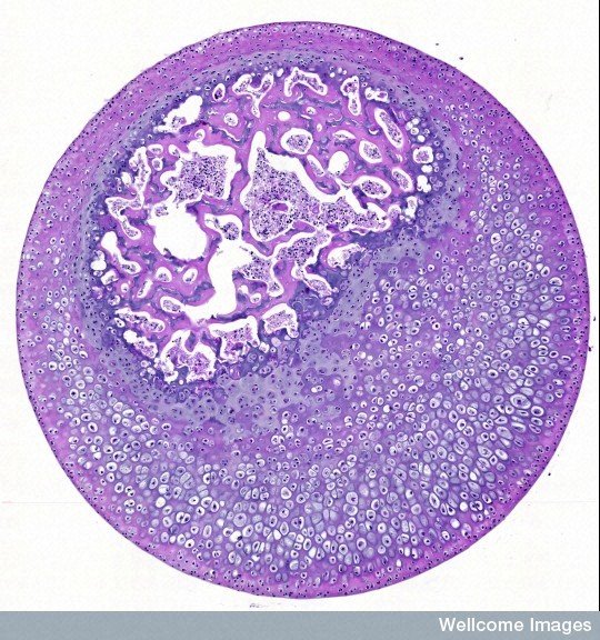
Identify the growth zone with reference to the diagrams above. Also refer to slide XX.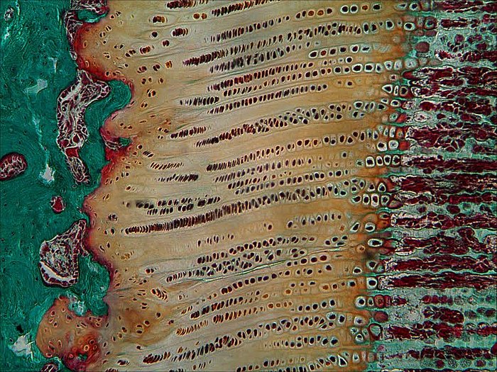
|
|